


By Dr. Robert Lowe
The following case demonstrates how to place a minimally invasive restoration using the caries detection mode of an intraoral camera and ACTIVA BioACTIVE-RESTORATIVE.
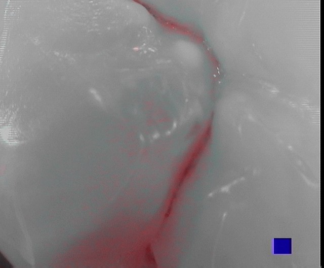
Figure 1 is an occlusal view of tooth number 29 taken with caries detection mode of an intraoral camera (SoproCare: Acteon USA) showing active caries in the central fissure of the tooth. It is important to note that this lesion is not yet able to “stick” with an explorer because of the depth of this narrow fissure and inability of the explorer tip to reach the lesion due to its larger size. A fissurotomy bur (micro NTF 6066: SS White) is used to carefully eradicate only the area that fluoresced positive (looks clinically like stain).
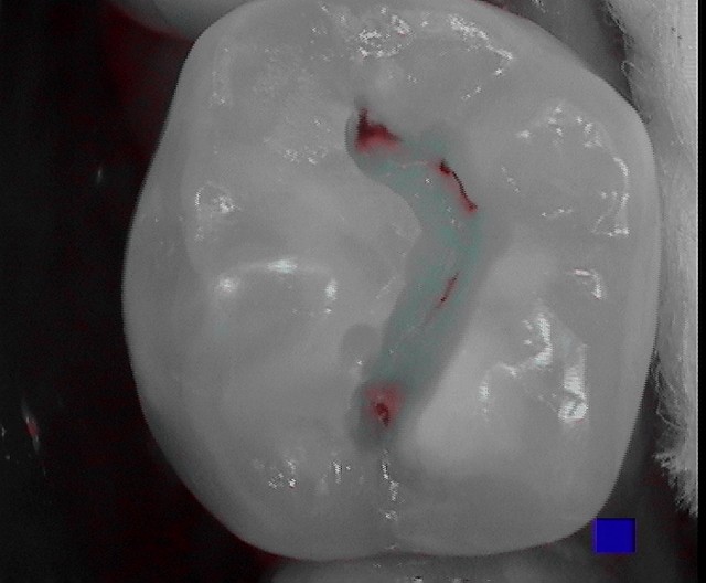
As the preparation is made, the intraoral camera on caries detection (Cario) mode can be used to check for complete active caries removal (Figure 2).
Because ACTIVA supports the natural remineralization process with release of calcium and phosphate, “white” areas of decalcified enamel can be preserved rather than removed during preparation. After using a total etch protocol, adhesive placement and light curing, ACTIVA BIOACTIVE-RESTORATIVE is placed into the micro-preparation using its automix tip with a bendable cannula (Figure 3).
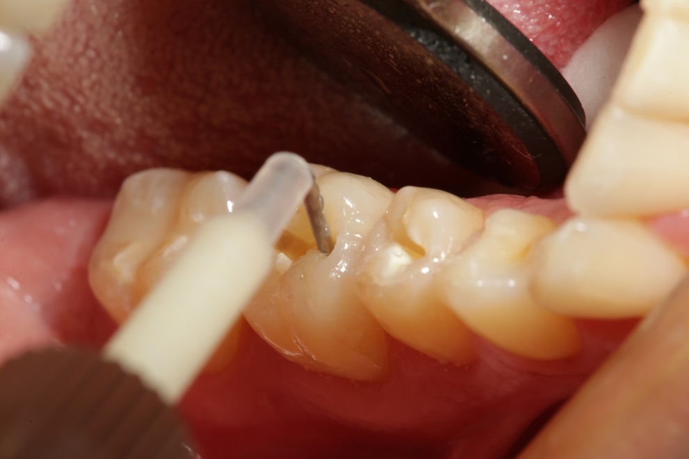
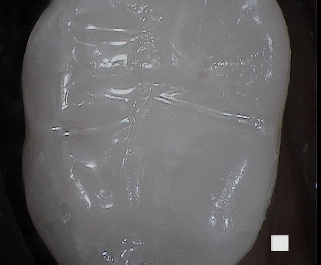
Figure 4 shows a view of the completed restoration taken with the intraoral camera. Note that it is difficult if not impossible to detect the restoration even at extreme magnification. Restorative margins are imperceptible. The tooth looks untouched like virgin enamel.
References
- Ferracane JL, Pfeifer CS, Bertassoni LE, “Biomaterials for Oral Health”, Dental Clinics of North America, vol.61, no.4, October 2017, pp. 651-872.
- Chao W, et al. Deflection at break of restorative materials. J Dent Res 94 (Spec Iss A) 2375, 2015 (iadr.org).
- Slowikowski L, et al. Fluoride ion release and recharge over time in three restoratives. Paper presented at: AADR Annual Meeting & Exhibition 2014; March 19, 2014; Boston, MA.
- Garcia-Gadoy F, Morrow BR, Pameijer CH, “Flexural Strength and Fatique of Activa RMGICs”, College of Dentistry UTHSC, Memphis and UConn School of Dentistry, Farmington, CT, White Paper Presentation at IADR, 2014.
- Maj J, Merritt J, Ferracane J, “Adhesion of S. Mutans Biofilms on Potentially Antimicrobial Dental Composites”, J dent Res 96 (Special Issue A); 2560, 2017.
- Comba A, Breschi L, et.al., J Dent Res 97 (Special Issue A) 0273, 2018.
- Girn VS, et.al., J Res Dent 93 (Special Issue A): 1163, 2014.

About Robert A. Lowe, DDS
Robert A. Lowe, DDS, received his Doctor of Dental Surgery degree from Loyola University School of Dentistry. After completing his residency, Dr. Lowe went into private practice and began to pursue another passion: clinical teaching. While running his own practice, Dr. Lowe served as a Clinical Professor in Restorative Dentistry at Loyola University School of Dentistry until its closure in 1993. In 2000, he relocated to Charlotte, NC.